
Venous thromboembolism (VTE)
Introduction to venous thromboembolism
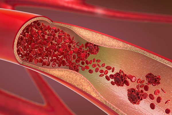
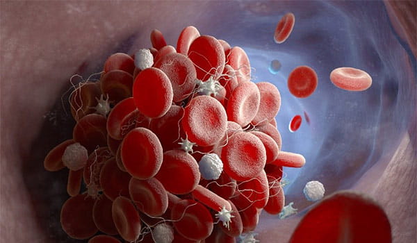
The venous system is the section of the circulatory system that uses veins to return the deoxygenated blood to the heart and lungs. Occasionally, irregularities in the wall of a vein especially in areas of slow flow, can cause a blood clot, or thrombus, to form [1].
VTE is an underdiagnosed preventable medical condition that can cause disability and death [2]. VTE includes deep vein thrombosis (DVT) and pulmonary embolism (PE).
Click here to renew consent
Deep vein thrombosis
DVT is preventable and treatable if discovered early [2].
Pulmonary embolism
The most serious complication of DVT happens when a part of the clot breaks off and travels through the bloodstream to the lungs, causing a blockage called pulmonary embolism (PE).
If the clot is small, and with appropriate treatment, people can recover from PE. However, there could be some damage to the lungs. If the clot is large, it can stop blood from reaching the lungs and is fatal [2].

Facts
- Every 37 seconds someone in the western world dies from a venous thromboembolism (VTE) [3]
- 1 in 4 people die from causes related to blood clots [3]
- 55%-60% of VTE cases occur during or following hospitalization [3]
- VTE is the #1 cause of preventable deaths in hospital [3]
- More people die from blood clots each year than the total number of people who lose their lives annually due to AIDS, breast cancer, and motor vehicle crashes combined [3]
There are a variety of risk factors that contribute to the development of deep vein thrombosis:
- Surgery, particularly surgery of the hip or leg, or abdominal surgery [4]
- Trauma or bone fracture [4]
- A long period of bed rest or sitting for a long time (e.g., on an airplane or in a car) [4]
- Cancer [4]
- Pregnancy [4]
- Birth control pills or hormones taken for symptoms of menopause [4]
- COVID-19: 7-39% of patients with COVID-19 infection who require mechanical ventilation have acute PE/DVT [5].
Diagnosis
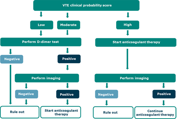
Diagnostic strategies for VTE are based on assessment of the pretest probability for individual patients, which provides an estimate of the expected prevalence of VTE at a population level [6].
For patients at low (unlikely) or intermediate VTE risk, using D-dimer* as the initial test reduces the need for diagnostic imaging [7].
For patients at high (likely) VTE risk, imaging is warranted.
For PE diagnosis, ventilation-perfusion scanning, and computed tomography pulmonary angiography (CTPA) are the most validated tests, whereas lower or upper extremity DVT diagnosis uses ultrasonography (ultrasound) [6].
* in combination with clinical probability assessment
References
2. Centers for Disease Control and Prevention, www.cdc.gov/ncbddd/dvt/facts.html Accessed Oct 2020
3. Thrombosis UK, www.thrombosisuk.org/Accessed Oct 2020
4. Stanford healthcare. www.stanfordhealthcare.org. Accessed Oct 2020
5. American Society of Hematology. https://www.hematology.org Accessed Oct 2020
6. Lim W et al. American Society of Hematology 2018 guidelines for management of venous thromboembolism: diagnosis of venous thromboembolism. Blood Adv. 2018; 27,22.
7. Konstantinides S et al. Guidelines for the diagnosis and management of acute pulmonary embolism developed in collaboration with the European Respiratory Society (ERS), Eur Heart J l 2019.
8. Strandberg K. The clinical use of -dimer assay, acutecaretesting.org, June 2017
Ez az oldal sütiket használ
Sütik használataIndtast en gyldig e-mail adresse
Vi vil snarest muligt sende dig en e-mailinvitation til at logge på med Microsoft Azure AD
Det ser ikke ud til, at din e-mail er registreret hos os
Venligst klik på "Kom i gang" knappen i e-mailen for at acceptere invitationen
Radiometer bruger Microsoft AZURE Active Directory (AZURE AD) til at bekræfte brugernes identitet.
Radiometer bruger AZURE AD til at give sikker adgang til dokumenter, ressourcer og andre tjenester på vores kundeportal for kunder og partnere.
Hvis din organisation allerede bruger AZURE AD, kan du bruge de samme identitetsoplysninger til at få adgang til Radiometers kundeportal.
Dine fordele
- Tillader brug af eksisterende Active Directory-identitetsoplysninger
- Enkel logon-oplevelse
- Brug samme identitetsoplysninger til at tilgå fremtidige tjenester
Anmod om adgang
Du vil via e-mail få en invitation til at tilgå vores tjenester, når din anmodning af blevet godkendt.
Når du accepterer invitationen, og hvis din organisation allerede bruger AZURE AD, kan du bruge de samme identitetsoplysninger til at få adgang til Radiometers kundeportal. Ellers vil du modtage et éengangs password, som bliver sendt til din e-mailadresse.